Cervical Disc Arthroplasty (CDA) is a relatively new procedure that is used to treat Disc Associated Wobbler's Syndrome (DAWS). It was developed by Dr. Filippo Adamo ECVN, the inventor of the artificial disc implant that we use in the procedure (The Adamo Spinal Disc). Dr. Adamo created the technique based on disc replacement surgeries that are done in people because of the tendency for DAWS patients to develop a 'domino lesion' of DAWS in the adjacent discs shortly after surgical correction of the first lesion.
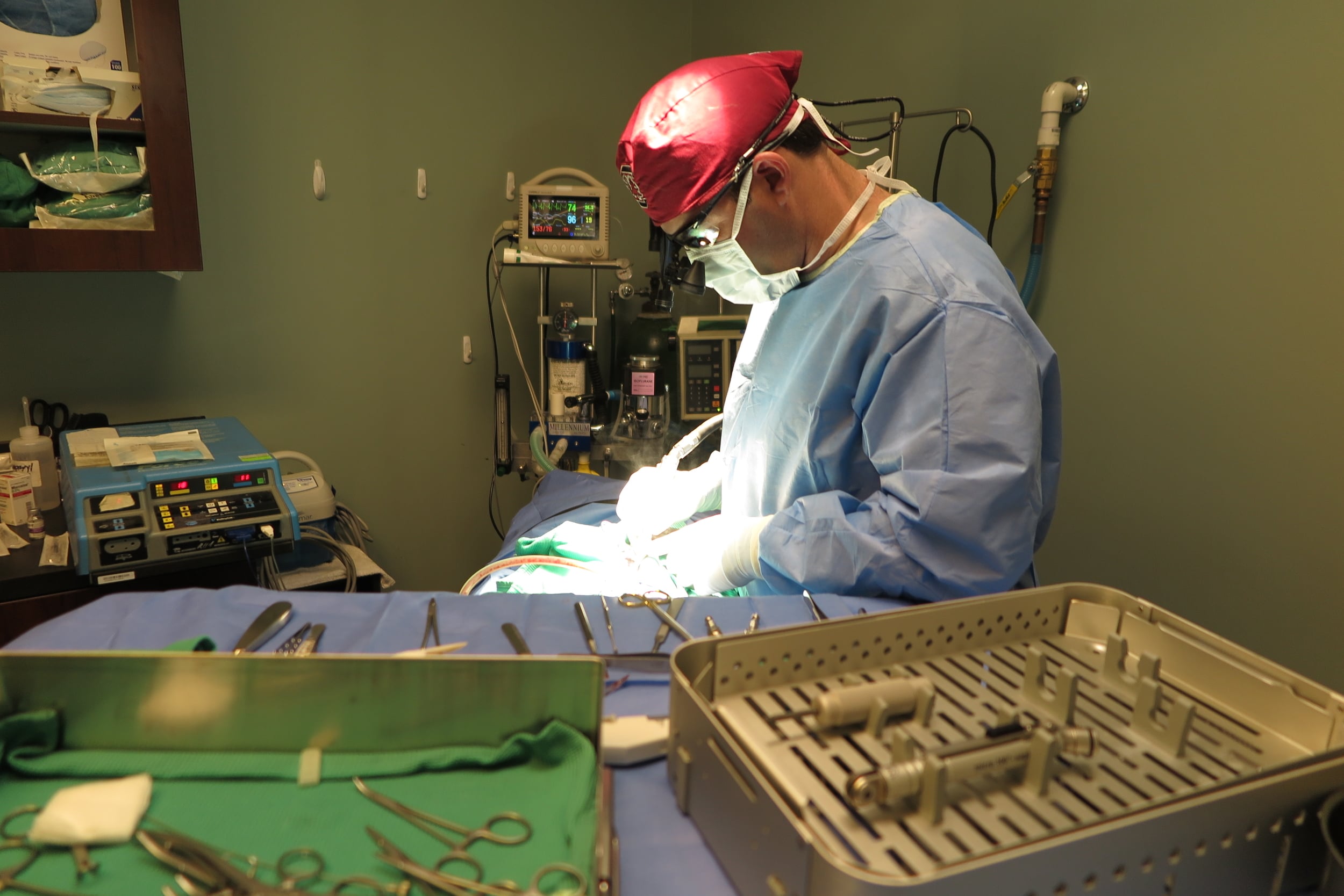
This is a picture of the instrument table and set-up before placing the disc implant in a patient.
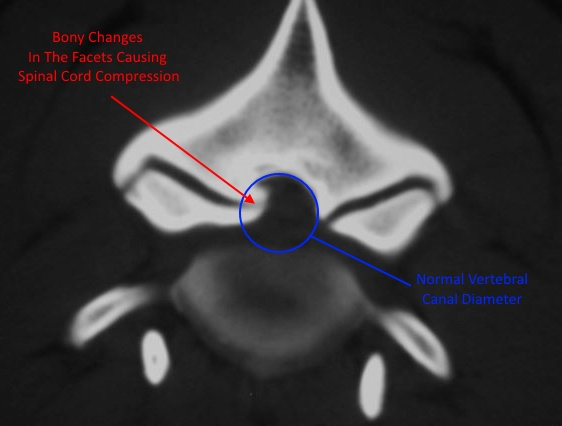
The disc implant serves two purposes in treating DAWS. First, it distracts the intervertebral space to relieve the compression on the spinal cord. This is similar to the stated goal-of-therapy of several of the older procedures as well. But where the CDA procedure differs, is in it's ability to maintain movement and range-of-motion at the surgical site. By maintaining the dynamic nature of the intervertebral disc space, the CDA technique seeks to minimize the effects on the adjacent discs and prevent the dreaded 'domino lesion'.

We can place more than one disc implant in a patient when indicated.
The Adamo Spinal Disc is currently in it's 5th iteration. The current design uses state-of-the-art manufacturing techniques and materials to improve the device and the surgical outcome for our patients.
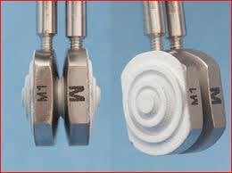
This is the complete Adamo Spinal Disc implant. The white material on the back of the implants promotes bony fusion between the vertebrae and the implant.
First, the device is made of surgical grade titanium. This lightweight and durable metal is also MRI safe. Which means that we can safely repeat MRI studies of the patients if needed after the surgery. In fact, repeat MRI studies are part of our post-operative evaluation for this procedure. Because the technique is so new and cutting edge, LOVN performs these post-operative MRI studies for these patients at no cost in exchange for using their outcomes and data for future studies and scientific papers. That's how much we believe in this procedure!
Second, the device uses a small thermoplastic articular surface to maintain the movement at the site. It works just like a 'ball and socket' to allow the two parts of the implant to move relative to one another. If the contact surfaces were made of metal, small microparticles would be produced from the friction and could lead to worsened inflammation or other problems.

Here you can see the two parts of the Adamo Spinal Disc and the 'ball and socket' articulation between the two. You can also see the screw threads that allow us to remove the positioning pins once the implant is in place.
Finally, the surface where the implant contacts the end plate bone of the vertebrae is specially treated in two ways to help in the healing process. First, the surface is treated in a very special way to create titanium 'pores' or tunnels that encourage bone growth into the implant. Once the bone grows into these 'tunnels' the interface between the bone and the implant becomes incredibly strong and durable. Secondly, the titanium porosities are coated with hydroxyapatite crystals. This is the same material that your body produces to create bone. It serves as a substrate to speed up the bone growth into the pores and the ultimate incorporation into the vertebrae.