This blog post is going to discuss the second form of "Wobbler's Disease" in dogs, Caudal Cervical Spondylomyelopathy or CCSM.
The breeds most commonly affected with CCSM include the Mastiffs and the Great Dane, but almost any large breed dog can be affected. Typically, this condition is seen in younger dogs as it is a developmental problem (contrasted with the degenerative problem that causes Disc Associated Wobbler's Disease or DAWS).
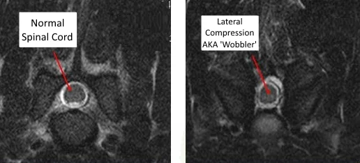
Again, to contrast with DAWS, the compression of the spinal cord is due to bony changes rather than discs, tendons, ligaments, or soft tissues. The parts of the vertebrae that form the bony tunnel of the vertebral canal develop abnormally and are thickened towards the tail-end of the vertebrae. This bony thickening is most noticable along the lateral (side) walls of the tunnel called the pedicles. So, the compression is from the side in CCSM, whereas it's mostly top-to-bottom with DAWS.
Once again, there are two different options for therapy, medical management vs surgery.
With medical management, the goal is to control the dog's clinical signs. But the drugs don't do anything to stop the disease progression. And, sometimes, the disease can progress very suddenly due to vascular or 'bruising' injuries to the spinal cord! Othertimes, the progression is slow and due to progressive bony changes at the true joints in the vertebrae, the facets.
Thickening of the facets can often cause progressive signs in CCSM due to worsening spinal cord compression...
Surgery attempts to alleviate the compression and works with medical management to control the clinical signs but also to halt the progression of the disease. In the ideal situation, surgical decompression even allows us to stop the medications.
Unlike DAWS which can suffer a 'domino effect' following surgery, this isn't common with CCSM decompression. But, just like DAWS, there are several surgical options for pets with CCSM. Some include decompression by removing bone from the top and sides of the vertebral canal. Others involve a distraction-fusion technique to allow the bone to resorb on its own and alleviate the compression.
Diagnosis of this disease often requires an MRI or a CT scan combined with a myelogram. In the end, the diagnosis will play an integral role in deciding which medical or surgical option is right for each individual patient.


