diagnostic testing

What is Canine Cognitive Dysfunction?

Did you know that dogs can develop a condition that is startlingly similar to Alzheimer's Disease in people?  It's true.  It's called Canine Cognitive Dysfunction (CCD) and a large percentage of our best friends will begin to show signs of this condition as early as 6 years old!  

Diagnosis of CCD is difficult.  There are sometimes changes on an MRI, but at other times all the diagnostic tests are normal.  That's why we, as veterinarians, rely so much on you, the pet owner, to identify signs of this condition and begin to intervene as soon as we can.  

You see, just like with Alzheimer's Disease, there is no cure for CCD.  There are lots of supplements, vitamins, botanical products, and even medications that can be used to slow the progression of the disease, but no one knows how to reverse the effects.

This particular disease is one that has caused me very personal heartbreak.  I had to say goodbye to the best-dang-dog-I've-ever-known, Scout, because of it.  

Professor Stout Scout in his prime ...

Professor Stout Scout in his prime ...

I met Professor Stout Scout when I was a technician working at an emergency veterinary clinic in Charleston, SC.  He came in the clinic as a stray and left that same night as my best friend and constant companion.  He went with me to Athens, GA for vet school; to Blacksburg, VA for my internship; and all the way to Davis, CA for my residency.

It was in California that I first started to notice some of the telltale signs of CCD.  Veterinarians use the acronym D-I-S-H to describe these signs.  

Disorientation is common.  

Interactions with their owners or other pets in the household may change

Sleep/Wake cycles are often disturbed

House-training can be lost as well

Scout started to show all these signs, and it seemed to be progressing rather rapidly.  Well, I'm insatiably curious and I had to know if my buddy was starting to show signs of CCD, or if there was something else going on.  Because brain tumors and other diseases can cause these signs too.  So Scout had an MRI and a spinal tap at UC Davis.

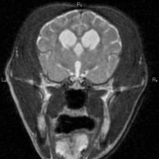
Typical structural changes in a dog's brain with CCD on MRI include loss of brain tissue, dilation of the fluid-filled ventricles, and shrinkage of the interthalamic adhesion.

Typical structural changes in a dog's brain with CCD on MRI include loss of brain tissue, dilation of the fluid-filled ventricles, and shrinkage of the interthalamic adhesion.

He must have read the same text books I was studying at the time, because he even had the structural brain changes that happen with CCD.  So, he'd checked all the tick-boxes for me.  I went on a search for something that might help.

I tried everything, and in the end found a cocktail of supplements and vitamins that seemed to slow his decline.  I was able to get another couple of years or so with him, but CCD won in the end.  Scout's quality of life declined, and I finally had to make that difficult, heartrending, and compassionate decision to help him slip off this mortal coil with all of the dignity and peace that I could muster for him.

So, if you think your older pet is starting to show signs of CCD, contact your veterinarian to have a discussion about the signs you're seeing.  We still can't reverse the signs of this disease so early intervention is key...

What is Chiari Malformation (COMS)?

Chiari malformation is the term used in human medicine that equates in veterinary medicine to Caudal Occipital Malformation Syndrome, COMS.  The most commonly affected breed with COMS is the Cavalier King Charles Spaniel.  With the advent of MRI we are finding this condition in several other breeds as well such as the Chihuahua, Yorkshire Terrier, and other small breeds of dogs.  

COMS is a condition where the base of the skull in the dog is abnormally shaped.  This leads to a more 'rounded skull' and, in turn, causes the part of the brain at the base of the skull (the cerebellum) to be 'crowded'.  Because this skull defect is present at birth and through the growth phase of the puppy, once the dog reaches adulthood, the skull changes are static and don't change.  But the after-effects are progressive.

This is a sagittal MRI of a dog with COMS.  You can see the cerebellum at the base of the skull has slipped out of the base of the skull into the 1st vertebrae of the neck in what is termed a cerebellar herniation.  The SM secondary to the…

This is a sagittal MRI of a dog with COMS.  You can see the cerebellum at the base of the skull has slipped out of the base of the skull into the 1st vertebrae of the neck in what is termed a cerebellar herniation.  The SM secondary to the COMS is present as the white area in the spinal cord just behind the cerebellum.

You see, COMS is associated with another condition that takes place in the spinal cord 'downstream' from the skull in the dog's neck.  This condition is called syringohydromyelia or SM.  SM is a progressive degenerative condition in the spinal cord that can happen anytime the flow of spinal fluid is disrupted.  

Scoliosis, or a left-right bending malformation f the spinal cord can also happen in cases of COMS and SM

Scoliosis, or a left-right bending malformation f the spinal cord can also happen in cases of COMS and SM

Most people know that the spinal cord has a left and right side that correspond directly to the left and right sides of the body.  If you damage the left side of the spinal cord, the signs associated with that damage will be seen on the left side of the body.  However, did you know that the spinal cord also has a 'front' and 'back' that carry specific types of information?  

The part of the spinal cord that is towards the chest of a person or animal carries information from the brain to the body, instructions on how to move.  The part of the spinal cord towards the back of the person or animal carries information from the body to the brain, sensory information if you will.

Interestingly enough, SM almost always forms in the sensory part of the spinal cord.  So it leads to abnormal sensations rather than weakness or abnormal movments.  That means that dogs with SM manifest some very odd and quite varied behaviors secondary to the abnormal sensations that their spinal cord is causing them to feel.  Imagine, that every time you got excited your toes began to itch and burn, your ear began to feel hot, or your arm became numb and tingly...

The most striking manifestation of this abnormal sensation is called 'phantom scratching'.  In this situation, dogs will begin to scratch 'at' their ear, but just a little bit off to the side.  Almost as if they were scratching the air!

Diagnosis of COMS and SM requires an MRI.  No other tests are able to definitively diagnose this condition.  There are other tests that have been used to support the diagnosis though, including BAER hearing tests, CT scans, ultrasound, and even infrared imaging.  

Treatment for COMS is sometimes medical, and sometimes surgical.  There are several drugs that can be used to help control the condition, and surgery can be very successful (depending on how one defines 'success').

What does the 'strength' of the MRI magnet mean?

MRI magnet strength is measured in a scientific unit called a Tesla.  For short-hand we use the abbreviation 'T' for this.  So a 1.5T magnet is one-and-a-half-times stronger than a 1.0T magnet.  There are MRI's used in experimental research that are 17.0T as of 2014 and there are even stronger magnetic fields that have been created!  The earth's magnetic field at the equator is only about 3 micro-Tesla by comparison.

So what does this little crash-course in electromagnetism mean for my pet?  

Well, the strength of the magnet is one of the more important variables in MR imaging.  It has a major impact on the resolution of the image and the speed with which we're able to acquire that image.  For comparison sake: with our 1.5T magnet we are able to scan the brain in a domestic feline in less than 20 minutes with excellent resolution and detail.  Previously, I've worked with a 1.0T magnet that would take closer to an hour and the resolution wouldn't be nearly as good.

Sagittal image of a dog's brain at 1.5T

Of course, there are other variables that can be adjusted to gain better resolution in a weaker magnet, but it takes a longer time.  And, when anesthesia is involved, less time is always safer!  Also, one has to be careful to consider the computer post-processing that goes into making these weaker magnet's images look good.  Just like Photo-shop can make anyone look like a super-slim super-model; images from these weaker magnets can be sharpened and manipulated to make them look as good as those from the stronger magnets.  But this can lead to false results when we try to interpret them.  It's a little like trying to gauge what's real when looking at the cover of a magazine!

What is an EEG?

EEG is the acronym for electroencephalography.  

Which is definitely a word that needs an acronym ....

Electroencephalography is a method of using electrodes that can sense the flow of electricity within biological tissues to look at brain function.  The brain, spinal cord, heart, and some other special tissues in the body use electrical signals to communicate rapidly between the cells that make up those particular tissues.  With these special electrodes, we can watch this electrical current and deduce information from them with regard to certain diseases.  

The machine shows us the electrical activity as several lines on a screen.  In animals and people, EEG is primarily used to evaluate an individual for seizures or, sometimes, sleep disorders.  

Seizures look like 'spikes' followed by 'waves' as you can see here...

Seizures look like 'spikes' followed by 'waves' as you can see here...

We also use it for patients that are in status epilepticus.  This is a condition when a seizure lasts longer than 20 minutes or there are multiple seizures without a return to consciousness in between.  Left untreated, this can cause permanent brain damage!

We've actually had two patients at LOVN this year that were in status epilepticus when they came in, but weren't having any outward signs.  This is called non-convulsive status epilepticus and without our EEG we wouldn't have been able to recognize it.  What's more, we were able to treat both of these patients with medications and use the EEG to monitor the effect of the drugs.

When patients are having such prolonged seizures, we often have to put them under general anesthesia to stop the seizures.  Then we have to decide when to try and wake them up.  Without the EEG this would just be a guess.  But with our EEG we can monitor the read-out for continued seizures even though the patient is completely asleep.  So, now, we don't have to guess when it's safe to wake a patient up.  We know...