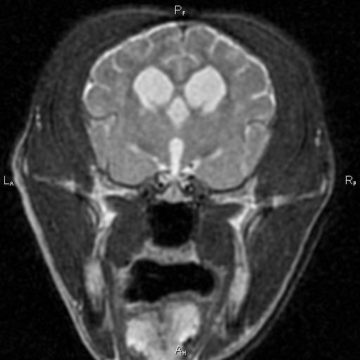
MRI

What is Canine Cognitive Dysfunction?

Did you know that dogs can develop a condition that is startlingly similar to Alzheimer's Disease in people?  It's true.  It's called Canine Cognitive Dysfunction (CCD) and a large percentage of our best friends will begin to show signs of this condition as early as 6 years old!  

Diagnosis of CCD is difficult.  There are sometimes changes on an MRI, but at other times all the diagnostic tests are normal.  That's why we, as veterinarians, rely so much on you, the pet owner, to identify signs of this condition and begin to intervene as soon as we can.  

You see, just like with Alzheimer's Disease, there is no cure for CCD.  There are lots of supplements, vitamins, botanical products, and even medications that can be used to slow the progression of the disease, but no one knows how to reverse the effects.

This particular disease is one that has caused me very personal heartbreak.  I had to say goodbye to the best-dang-dog-I've-ever-known, Scout, because of it.  

Professor Stout Scout in his prime ...

Professor Stout Scout in his prime ...

I met Professor Stout Scout when I was a technician working at an emergency veterinary clinic in Charleston, SC.  He came in the clinic as a stray and left that same night as my best friend and constant companion.  He went with me to Athens, GA for vet school; to Blacksburg, VA for my internship; and all the way to Davis, CA for my residency.

It was in California that I first started to notice some of the telltale signs of CCD.  Veterinarians use the acronym D-I-S-H to describe these signs.  

Disorientation is common.  

Interactions with their owners or other pets in the household may change

Sleep/Wake cycles are often disturbed

House-training can be lost as well

Scout started to show all these signs, and it seemed to be progressing rather rapidly.  Well, I'm insatiably curious and I had to know if my buddy was starting to show signs of CCD, or if there was something else going on.  Because brain tumors and other diseases can cause these signs too.  So Scout had an MRI and a spinal tap at UC Davis.

Typical structural changes in a dog's brain with CCD on MRI include loss of brain tissue, dilation of the fluid-filled ventricles, and shrinkage of the interthalamic adhesion.

Typical structural changes in a dog's brain with CCD on MRI include loss of brain tissue, dilation of the fluid-filled ventricles, and shrinkage of the interthalamic adhesion.

He must have read the same text books I was studying at the time, because he even had the structural brain changes that happen with CCD.  So, he'd checked all the tick-boxes for me.  I went on a search for something that might help.

I tried everything, and in the end found a cocktail of supplements and vitamins that seemed to slow his decline.  I was able to get another couple of years or so with him, but CCD won in the end.  Scout's quality of life declined, and I finally had to make that difficult, heartrending, and compassionate decision to help him slip off this mortal coil with all of the dignity and peace that I could muster for him.

So, if you think your older pet is starting to show signs of CCD, contact your veterinarian to have a discussion about the signs you're seeing.  We still can't reverse the signs of this disease so early intervention is key...

What is the Treatment for Chiari-like Malformation (COMS)?

I often describe the disease process involved in these cases using the analogy of putting your finger over the end of a garden hose.  When you do, the same volume of water comes through as it did without your finger in place, but the aperture is much smaller.  This causes changes in the hydrostatic pressure of the flow through the end of the garden hose.  

With every heartbeat, there is a sudden surge of fluid through the hole at the base of the skull.  This allows the skull to accommodate the increased volume of blood that comes in with every heartbeat. But the change in pressure that happens with the cerebellum herniated (or just crowded) at the base of the skull is just like having your thumb over the end of the garden hose ...

This is a video of a special MRI sequence performed in a human that shows the pulsatile flow of spinal fluid caused by the heartbeat ...

Management of this condition with medications (like prednisone and omeprazole) attempts to reduce the production of spinal fluid.  This is analogous to turning down the spigot while keeping your thumb over the end of the garden hose ...


Surgery removes the compression on the cerebellum to allow the fluid to pass through more easily.  This is akin to taking your thumb off the garden hose ...


I mention all of this, because I want to remind everyone that surgery doesn't directly make the syringohydromyelia (SM) in the spinal cord go away.  Rather, it prevents the disease from getting worse by removing the underlying cause.  That's important because it plays into how we define 'success' when looking at surgery.

I always caution my clients to think of a successful surgery as - a patient that recovers from the procedure with no complications and does not get worse over time due to progression of the SM.  In other words, these patients may still require medications and the surgery does not (necessarily) make the SM go away.  If we get improvement beyond this definition of success, then I call that 'icing on the cake'.  Using this definition of success, we are successful with surgery more than 80% of the time.  

Most of the surgical failures happen months to years after the procedure due to scar formation at the surgical site.  This seems to be where the titanium mesh has its greatest benefit.  Since I've been using the mesh, I haven't had any patients worsen in that time frame.  

Also, when we remove the bone, there is still a thick membrane (the meninges and particularly the dura mater) that restricts and confines the cerebellum and brain.  By patching in a piece of tissue, we can make more room within this membrane and relieve the compression.  It's very similar to letting out the waist in a pair of pants.  To help prevent scar tissue from forming here and undoing all that we've done, I use tissue collected from the patient's themselves (autograft) rather than material that might cause more inflammation or rejection of the graft.

What does the 'strength' of the MRI magnet mean?

MRI magnet strength is measured in a scientific unit called a Tesla.  For short-hand we use the abbreviation 'T' for this.  So a 1.5T magnet is one-and-a-half-times stronger than a 1.0T magnet.  There are MRI's used in experimental research that are 17.0T as of 2014 and there are even stronger magnetic fields that have been created!  The earth's magnetic field at the equator is only about 3 micro-Tesla by comparison.

So what does this little crash-course in electromagnetism mean for my pet?  

Well, the strength of the magnet is one of the more important variables in MR imaging.  It has a major impact on the resolution of the image and the speed with which we're able to acquire that image.  For comparison sake: with our 1.5T magnet we are able to scan the brain in a domestic feline in less than 20 minutes with excellent resolution and detail.  Previously, I've worked with a 1.0T magnet that would take closer to an hour and the resolution wouldn't be nearly as good.

Sagittal image of a dog's brain at 1.5T

Of course, there are other variables that can be adjusted to gain better resolution in a weaker magnet, but it takes a longer time.  And, when anesthesia is involved, less time is always safer!  Also, one has to be careful to consider the computer post-processing that goes into making these weaker magnet's images look good.  Just like Photo-shop can make anyone look like a super-slim super-model; images from these weaker magnets can be sharpened and manipulated to make them look as good as those from the stronger magnets.  But this can lead to false results when we try to interpret them.  It's a little like trying to gauge what's real when looking at the cover of a magazine!

What is Magnetic Resonance Imaging?

Magnetic Resonance Imaging, or MRI, has been used since the late 1970's to image the human body.  Among the different ways that doctors have to image ourselves and our animal friends, the MRI is unique.  Unlike an x-ray, CAT scan, PET scan, or other radiologic techniques, the MRI doesn't use radiation to create the images.  Rather it relies on the magnetic properties of the tissues in our bodies.

When a patient is placed in the MRI machine, that individual is subjected to a strong magnetic field.  The MRI machine then pulses radiowaves into the part of the body we are inspecting and 'listens' for the return radiowaves the tissue gives off.  This lets us determine specific characteristics of the tissues we're looking at and gain unprecedented insight into the architecture and disease state of the body.  When we give an intravenous injection of contrast, this improves our ability to recognize and characterize the disease even more.

As MRI technology has improved, doctors have been able to find even more detail and have even begun to be able to see the functional activity of parts of the brain.  But some of the most exciting research is being done with diffusion-weighted imaging.  This technique allows us to distinguish 'strokes' from tumors and other diseases.  But in some of the very advanced MRI machines used in research, scientists are able to track the connections between parts of the brain and begin to understand the 'wiring' that makes us all the amazing manifestations of Life that we are.

Check out the Human Connectome Project for some amazing images!