Chiari malformation

What is the Treatment for Chiari-like Malformation (COMS)?

I often describe the disease process involved in these cases using the analogy of putting your finger over the end of a garden hose.  When you do, the same volume of water comes through as it did without your finger in place, but the aperture is much smaller.  This causes changes in the hydrostatic pressure of the flow through the end of the garden hose.  

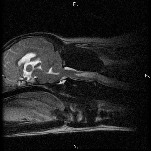
With every heartbeat, there is a sudden surge of fluid through the hole at the base of the skull.  This allows the skull to accommodate the increased volume of blood that comes in with every heartbeat. But the change in pressure that happens with the cerebellum herniated (or just crowded) at the base of the skull is just like having your thumb over the end of the garden hose ...

This is a video of a special MRI sequence performed in a human that shows the pulsatile flow of spinal fluid caused by the heartbeat ...

Management of this condition with medications (like prednisone and omeprazole) attempts to reduce the production of spinal fluid.  This is analogous to turning down the spigot while keeping your thumb over the end of the garden hose ...


Surgery removes the compression on the cerebellum to allow the fluid to pass through more easily.  This is akin to taking your thumb off the garden hose ...


I mention all of this, because I want to remind everyone that surgery doesn't directly make the syringohydromyelia (SM) in the spinal cord go away.  Rather, it prevents the disease from getting worse by removing the underlying cause.  That's important because it plays into how we define 'success' when looking at surgery.

I always caution my clients to think of a successful surgery as - a patient that recovers from the procedure with no complications and does not get worse over time due to progression of the SM.  In other words, these patients may still require medications and the surgery does not (necessarily) make the SM go away.  If we get improvement beyond this definition of success, then I call that 'icing on the cake'.  Using this definition of success, we are successful with surgery more than 80% of the time.  

Most of the surgical failures happen months to years after the procedure due to scar formation at the surgical site.  This seems to be where the titanium mesh has its greatest benefit.  Since I've been using the mesh, I haven't had any patients worsen in that time frame.  

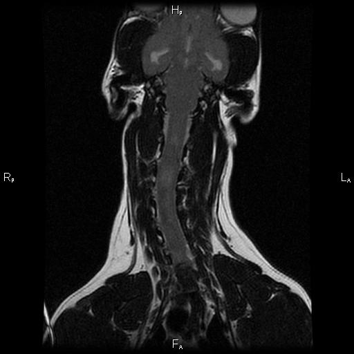
Also, when we remove the bone, there is still a thick membrane (the meninges and particularly the dura mater) that restricts and confines the cerebellum and brain.  By patching in a piece of tissue, we can make more room within this membrane and relieve the compression.  It's very similar to letting out the waist in a pair of pants.  To help prevent scar tissue from forming here and undoing all that we've done, I use tissue collected from the patient's themselves (autograft) rather than material that might cause more inflammation or rejection of the graft.

What is Chiari Malformation (COMS)?

Chiari malformation is the term used in human medicine that equates in veterinary medicine to Caudal Occipital Malformation Syndrome, COMS.  The most commonly affected breed with COMS is the Cavalier King Charles Spaniel.  With the advent of MRI we are finding this condition in several other breeds as well such as the Chihuahua, Yorkshire Terrier, and other small breeds of dogs.  

COMS is a condition where the base of the skull in the dog is abnormally shaped.  This leads to a more 'rounded skull' and, in turn, causes the part of the brain at the base of the skull (the cerebellum) to be 'crowded'.  Because this skull defect is present at birth and through the growth phase of the puppy, once the dog reaches adulthood, the skull changes are static and don't change.  But the after-effects are progressive.

This is a sagittal MRI of a dog with COMS.  You can see the cerebellum at the base of the skull has slipped out of the base of the skull into the 1st vertebrae of the neck in what is termed a cerebellar herniation.  The SM secondary to the…

This is a sagittal MRI of a dog with COMS.  You can see the cerebellum at the base of the skull has slipped out of the base of the skull into the 1st vertebrae of the neck in what is termed a cerebellar herniation.  The SM secondary to the COMS is present as the white area in the spinal cord just behind the cerebellum.

You see, COMS is associated with another condition that takes place in the spinal cord 'downstream' from the skull in the dog's neck.  This condition is called syringohydromyelia or SM.  SM is a progressive degenerative condition in the spinal cord that can happen anytime the flow of spinal fluid is disrupted.  

Scoliosis, or a left-right bending malformation f the spinal cord can also happen in cases of COMS and SM

Scoliosis, or a left-right bending malformation f the spinal cord can also happen in cases of COMS and SM

Most people know that the spinal cord has a left and right side that correspond directly to the left and right sides of the body.  If you damage the left side of the spinal cord, the signs associated with that damage will be seen on the left side of the body.  However, did you know that the spinal cord also has a 'front' and 'back' that carry specific types of information?  

The part of the spinal cord that is towards the chest of a person or animal carries information from the brain to the body, instructions on how to move.  The part of the spinal cord towards the back of the person or animal carries information from the body to the brain, sensory information if you will.

Interestingly enough, SM almost always forms in the sensory part of the spinal cord.  So it leads to abnormal sensations rather than weakness or abnormal movments.  That means that dogs with SM manifest some very odd and quite varied behaviors secondary to the abnormal sensations that their spinal cord is causing them to feel.  Imagine, that every time you got excited your toes began to itch and burn, your ear began to feel hot, or your arm became numb and tingly...

The most striking manifestation of this abnormal sensation is called 'phantom scratching'.  In this situation, dogs will begin to scratch 'at' their ear, but just a little bit off to the side.  Almost as if they were scratching the air!

Diagnosis of COMS and SM requires an MRI.  No other tests are able to definitively diagnose this condition.  There are other tests that have been used to support the diagnosis though, including BAER hearing tests, CT scans, ultrasound, and even infrared imaging.  

Treatment for COMS is sometimes medical, and sometimes surgical.  There are several drugs that can be used to help control the condition, and surgery can be very successful (depending on how one defines 'success').